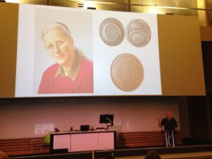
The Beddington Medal is the BSDB’s major commendation to promising young biologists, awarded for the best PhD thesis in Developmental Biology defended in the year previous to the award. Rosa Beddington was one of the greatest talents and inspirational leaders in the field of developmental biology. Rosa made an enormous contribution to the field in general and to the BSDB in particular, so it seemed entirely appropriate that the Society should establish a lasting memorial to her. The design of the medal, mice on a stylised DNA helix, is from artwork by Rosa herself. We would like to congratulate the 2019 winner of the Beddington Medal, David Munro, and would like to take this opportunity to give a brief overview of his career and the PhD project that was awarded the Beddington medal.
Jim Smith introduced the Beddington medal with heartfelt memories of Rosa Beddington and her time at the NIMR. Please read more of his thoughts here.
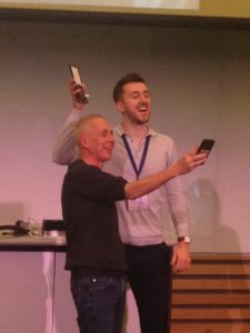
Some complicated selfies were taken as the medal was passed over before David went on to present the work that has deserved him this award. In the words of his PhD supervisor:
“The really impressive thing about David’s work is that he did not come to my lab to fit in with an existing line of research but created one of his own”. Jamie Davies, University of Edinburgh.
David received his undergraduate degree in Sport and Exercise Science at the University of Stirling (2010-2014). With this, he achieved a first-class honours degree and the prize for the best overall performance throughout a physiology related degree (British Physiological Society Undergraduate Prize). His dissertation project investigated associations between ADRB2 mutations (an adrenaline receptor gene in humans) and athlete status/athletic ability measurements. Subsequently, he was awarded a University of Stirling Head of School Summer Bursary Award to remain in Stirling during the summer of 2014 and investigate the relationship between transcribed ultra-conserved regions of RNA (T-UCRs) and the development of diet-induced insulin resistance in humans (Summer 2014). He then moved to the University of Edinburgh for his MSc by Research in Biomedical Sciences (2014-2015). Again, he received a distinction and was awarded the Class Prize for best student. During this time, he studied the physiology of S-acylation the regulation of skeletal muscle energy expenditure by an obesity-associated phospholipase as part of two research placements.
David has been awarded the Beddington medal for his exceptional work performed during his 3-year MRC-funded PhD at the University of Edinburgh with Prof Jamie Davies and Dr Peter Hohenstein (2015-2018): The thesis is titled ‘Mechanisms of kidney vascularisation and the roles of macrophages in renal organogenesis’. During his PhD, he gave several oral and poster presentations at national and international conferences, supervised students (including a Gurdon Summer Studentship Awardee), established numerous international collaborations, was awarded travel grants (including a BSDB Conference Grant), and reviewed manuscripts for leading journals (including Cell Reports, Angiogenesis, and Scientific Reports). He is now a post-doctoral fellow at the UK Dementia Research Institute (University of Edinburgh; 2019- present), continuing research in macrophage biology under the supervision of Prof Josef Priller. His current focus in on brain macrophages (microglia) in development, neurodegeneration, and aging.
Thesis description
Kidneys are specialised organs that clean the blood, removing waste while retaining what is useful. This requires a complex vasculature, and its formation as a foetus develops is poorly understood. I started my PhD research by using advanced microscopy techniques to visualise how blood vessels form in three-dimensions in the mouse kidney. In doing so, I identified when and from where the first blood vessels enter the kidney, and how blood vessels pattern at the edge of the kidney throughout development.
Blood vessels can form through angiogenesis (branching of new vessels from pre-existing ones) and/or vasculogenesis (assembly of new vessels from the coalescence of endothelial precursor cells). It has long been thought that a combination of both processes occurs during kidney vascularisation; however, my thesis work indicates that this concept may not be correct. My data instead suggest that kidney vascularization relies on growth and remodelling of pre-existing vessels (angiogenesis) and does not depend on vasculogenesis at any point (Publications 1 and 5 in CV). When assessing the entire 3D vascular tree of the kidney, isolated endothelial cells were never observed at any developmental age. Instead, all vessels, including the newly forming ones, were connected to pre-existing vessels that could be traced to the major circulatory vessels.
I then focused on the blood vessels at the edge of the kidney, which I found to consistently and accurately pattern around a special collection of cells – the cap mesenchyme. The cap mesenchyme contains cells that eventually become the cleaning tubes of the kidney, the nephrons. This cell population undergoes rounds of splitting at the kidney’s periphery. As this happens, I demonstrated that blood vessels migrate through the newly opened regions between the separating cap mesenchymal populations (Publication 1 in CV). This occurs in cycles throughout development and is likely to be vital for the oxygenation of the kidney’s outer region, the site where important processes such as nephron formation take place.
I determined that a signalling molecule, semaphorin-3f, and its receptor, neuropilin-2, were expressed in a pattern consistent with them having roles in this cyclical patterning of blood vessels; however, using mouse models where the genes for these molecules were deleted, I established that they were not vital for this process (Publication 2 in CV).
I next shifted my research focus towards a specialised cell type known as the macrophage (macro = big; phage = eater) in the developing kidney (Publication 3 in CV). Macrophages are immune cells best known for clearing foreign and damaged cells. These cells have vital roles during animal development, but little is known about their specific functions during kidney development.
Macrophages arrived in the mouse kidney early during its development, where they were required to clear away misplaced cells to ‘set-the-stage’ for early kidney development (Publication 6 in CV, under review). Throughout later development, most macrophages wrapped around blood vessels and I demonstrated their ability to eat endothelial cells (which usually line the blood vessels) and red blood cells (which are carried within them) within the kidney. I also established that kidney macrophages produced many molecules linked to blood vessel development, and so I examined the consequences of macrophage-loss on blood vessel formation. Blood vessels normally form continuous networks in the kidney; however, when macrophages were depleted (by blocking a macrophage-survival signalling pathway), connections between renal blood vessels were reduced (Publication 6 in CV).
Publications
Munro DAD, Hohenstein P, Davies JA. 2017. Cycles of vascular plexus formation within the nephrogenic zone of the developing kidney. Scientific Reports. 7: 3273.
Munro DAD, Hohenstein P, Coate TM, Davies JA. 2017. Refuting the hypothesis that semaphorin-3f/neuropilin-2 guide endothelial patterning around the cap mesenchyme in the developing kidney. Developmental Dynamics. 246:1047-1056.
Munro DAD, Hughes J. 2017. The Origins and Functions of Tissue-Resident Macrophages in Kidney Development. Frontiers in Physiology. 8:837. (Review)
Mills CG, Lawrence ML, Munro DAD, El-Hendawi M, Mullins JJ, Davies JA. 2017. Asymmetric BMP4 signalling improves the realism of kidney organoids. Scientific Reports. 7:14824.
Munro DAD, Davies JA. 2018. Vascularizing the kidney in the embryo and organoid: questioning assumptions about renal vasculogenesis. Journal of the American Society of Nephrology. (Perspectives article).
Munro DAD, et al. Macrophages restrict the nephrogenic field and promote endothelial connections during kidney development. eLife 2019;8:e43271 DOI: 10.7554/eLife.43271
Upon receiving a 4-year VIB International PhD Program grant, Mariya joined the lab of An Zwijsen in Leuven, Belgium to study the origins of amniotic stem cells and to dissect the unique extraembryonic defects of the Smad5 knock-out mouse embryos. SMAD5 is a downstream effector of BMP signaling, a major pathway involved in many processes in development and cancer. Mariya was fascinated by how entangled the development of embryonic and extraembryonic tissues during early development is, and appreciated the importance of understanding better these neglected parts of the conceptus. She contributed to the finding that Smad5 mutant embryos develop an ectopic primitive streak-like/tumor-like structure in their amnion due to defective signaling (Periera et al., 2012, Development 139(18)), and identified amnion-specific set of marker genes for mouse and human (Dobreva et al., 2012, Stem Cells Int. 987185). The culmination of Mariya’s PhD and postdoc work at Zwijsen’s lab was her most recent paper entitled “Amniotic ectoderm expansion in mouse occurs via distinct modes and requires SMAD5-mediated signalling” (Dobreva et al., 2018, Development 145(15)). This work impressed the judges of the Denis Summberbell Lecture award as a thorough study that sheds light upon both the origin of amnion and the molecular dynamics of its development combining cutting-edge, classical, and original techniques.
After a career brake, Mariya received a 2-year Marie Skłodowska-Curie fellowship and in 2016 moved to the UK to join the lab of Arkhat Abzhanov at Imperial College London. Expanding her research interests towards evolutionary developmental biology, she currently studies the developmental mechanisms underlying the rapid evolution and adaptive radiation of Darwin’s finches from Galapagos islands.
Lecture abstract:
Upon gastrulation, the mammalian conceptus transforms rapidly from a simple bilayer into a multi-layered embryo enveloped by its extraembryonic membranes. The embryonic-extraembryonic junction is a hot spot for dynamic cell rearrangements that drive early morphogenesis. The innermost extraembryonic membrane, the amnion, develops at the embryonic-extraembryonic interphase and gradually encases the developing conceptus. Impaired amnion development causes major embryonic malformations, yet its origin remains ill-defined. Mouse embryos, deficient in the BMP signalling effector SMAD5, show aberrant amnion and ventral folding morphogenesis and delayed closure of the proamniotic canal. I developed a microdissection technique and sequenced the transcriptomes of individual Smad5 mutant amnions isolated before the first visible malformations appear (E7.0-E7.5). I revealed two sets of defective amnions: one with a primitive-streak mesoderm signature and another one with unexpected chorionic ectoderm signature. Tetraploid chimera and immunostaining assays indicated that, in both cases, a deficit in the expansion of amniotic ectoderm results in inclusion of non-amniotic, non-squamous tissues in the amniotic microenvironment. Interestingly, the inclusions can be either of embryonic or of extraembryonic origin. To explain the different types of Smad5 mutant defects and to clarify the origin of mouse amnion, we related our findings to existing clonal analysis of early mouse embryos performed by Kirstie A. Lawson (University of Edinburgh). She traced the fate of single cells labeled before amnion formation. Four clone types contribute to the amniotic ectoderm with distinct growth patterns. Two main clone types were identified, with progenitors in the extreme proximal-anterior epiblast. Their early descendants initiate and expand amniotic ectoderm posteriorly, following the progression of the developing amniochorionic fold. Surprisingly, descendants of cells remaining anteriorly, later expand the amniotic ectoderm from its anterior side. The progenitor regions of all types are close to BMP sources in extraembryonic ectoderm and visceral endoderm. We attribute the two Smad5 mutant defect types to impairment of progenitors of the two main cell populations in amniotic ectoderm, and to compromised cuboidal-to-squamous transition of the anterior amniotic ectoderm. In both cases, SMAD5 is critical for expanding the amniotic ectoderm rapidly into a stretchable squamous sheet to accommodate exocoelom expansion, axial growth and folding morphogenesis.
This year, is the BSDB’s 70th anniversary, and this was clearly reflected at our Spring Meeting, 15-18 April 2018 in Warwick! Apart from an outstanding speaker list, and the award of most BSDB medals & prizes of 2018, we saw a very special event with many extras, as is well described here by Dillan Saunders. Dillan undertook his BSc Honours with Michael Akam studying centipedes, is currently performing his MSc in the lab of Megan Davey date-mapping the developing limb bud of chick with novel transgenic technologies. and will be returning to Cambridge later this year to begin The Wellcome Trust Developmental Biology PhD program. Dillan’s blog post aligns with a long-standing tradition of the BSDB to engage young members (see our archive blog), and we strongly encourage PhD students and postdocs to make their voice heard by writing reports or articles for our website and newsletter.
The British Society for Developmental Biology (BSDB) recently held its annual Spring Meeting at the University of Warwick. This was no ordinary meeting, though it is fair to say that BSDB meetings rarely are. This Spring Meeting celebrated the 70th anniversary of the BSDB and did so excellently, birthday cakes and all. With a retrospective look at the past of the society, and fantastic speakers showcasing present and ongoing work, the stage was set for a meeting that not only celebrated a strong history, but also looked forward, critically yet hopefully, to the future of Developmental Biology and of the BSDB itself.
The conference began with a career workshop for students and post-docs (see our blog post). In keeping with the celebration of the BSDB’s birthday, the focus of the workshop was on staying in academia. A variety of speakers and other group leaders fielded questions and shared details of the personal journeys to have brought them to their current positions. I had the opportunity to speak to Henrik Semb – whose example shows that not everyone follows a straight, let alone predictable, career trajectory. Judith Kimble shared her views on not seeing the lab as a place of work, and finally I got to hear Ottoline Leyser’s thoughts on work-life balance; a phrase which misleadingly implies that the two are opposed to one another. It would however be in our best interests, she explained, to see work and life as overlapping and complementary to one another.
The career workshop
After the careers workshop came the first plenary talk, in which Eric Wieschaus explained his recent work on mesoderm invagination in Drosophila and how one transcriptional activation can lead to a sequence of events (Weng and Wieschaus, 2017). Thematically paired with this talk was Maria Leptin’s plenary lecture, in which she discussed the development of in silico models for actin dynamics in order to recapitulate mesoderm invagination (Belmonte et al., 2017).
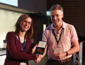
Emilia receives the Beddington medal from Simon Bullock
Following a short break, the Beddington Medal was awarded to Emily Favuzzi for the best PhD thesis in Developmental Biology of the year, which she performed on the transcriptional networks at play during interneuron development (see our blog post). She then gave a talk that illustrated how meticulous and comprehensive her work was (Favuzzi et al., 2017). This was followed up by a plenary talk from Marianne Bronner on the transcriptional networks of specific populations of neural crest cells, with attention to how her work with lampreys shows that the neural crest has acquired additional functions in the jawed vertebrates (Green et al., 2017). Unfazed by technical difficulties with the final slides of the presentation, Bronner took on the role of the lamprey and humorously indicated on herself the location of the neural crest cells.
The evening was capped off by three events that celebrated the history, and looked into the future, of the BSDB and Developmental Biology as a whole. First, came two talks from historians of science, Nick Hopwood and Tim Horder. Hopwood’s talk detailed the story of the crystallization of modern Developmental Biology in the late ‘40s and ‘50s. He described how the London Embryologists Club began in 1948 and how it then broadened both its geographical location and the field that it represented to form the BSDB (see also our archive blog).
Nick Hopwood’s lecture about DB’s history
Off the back of the historians’ view of the past, came a panel discussion of the future of the field. The panel was made up of Ottoline Leyser, James Briscoe, Maria Leptin, Jonathan Slack, Judith Kimble and Patrick Lemaire. They fielded several thought-provoking questions from the audience, which led to a lively discussion. As might be expected from a big anniversary meeting, there was much reminiscing on the early days of molecular Developmental Biology in the 1980s, often referred to as the ‘Golden Age.’ When asked what made this era such an exciting time for Developmental Biology, Jonathan Slack noted that it was a time when developmental biologists were becoming dissatisfied with the explanations of the previous heyday of embryology in the 1930s.
In the subsequent discussion, Ottoline Leyser pointed out that, what with the advent of new technologies and ideas, we are in fact in the midst of a golden age for Developmental Biology. Indeed, though there are still some who believe that certain areas of Developmental Biology hold no more secrets, recent years have shown new developments in, for example, the study of the anterior-posterior axis in Drosophila (Clark and Akam, 2016) and the C. elegans cell lineage (Sammut et al., 2015).
Other points of discussion included the importance of studying development within the context of time and the great potential of computational modelling. Discussed also were the logistics of maintaining an idea of the overall picture without becoming too focused on a single model system. The final event of the day was an informal round table discussion where the points raised previously, and many others, were discussed at length over much wine.
Monday began with a plenary talk from Matthew Freeman, amusingly titled ‘Confessions of an ex-developmental biologist’, in which he described his current focus on cell signalling and the pertinence of Cell Biology to the understanding of development (e.g. Christova et al., 2013). Following this, the two morning sessions covered ‘TISSUE AND ORGAN DEVELOPMENT’ and ‘DEVELOPMENTAL GENE REGULATORY NETWORKS.’ I listened to James Briscoe’s talk on the Sonic Hedgehog morphogen gradient in the neural tube and the interesting mathematical models that his lab has used to explore the time and precision of its patterning. Virginia Papaioannou then spoke about the role of Tbx6 in left-right axis establishment (Concepcion et al., 2017).
Also in this session were complementing talks from Eileen Furlong (Mikhaylichenko et al., 2018) and Mike Levine (Lim et al., 2018) on the relationships between enhancers and promoters and how chromatin architecture regulates gene expression. After a lunch poster session, the afternoon sessions began with talks on ‘MECHANISMS OF GLOBAL GENE REGULATION’ and ‘CELL BIOLOGY AND DEVELOPMENT’. That afternoon, I caught a talk by Robb Krumlauf, in which he showed some fascinating lamprey experiments which supplied further detail on the ancient interactions between retinoic acid and Hox genes. Following this, Caroline Telfer demonstrated her impressive quantity of PhD work on the upstream regulation of the GATA genes, and then Pavel Tomancak showed a combination of beautiful live imaging and computational models for serosa closure in Tribolium castaneum embryos. The day’s closing plenary lecture was given by Janet Rossant, which included her work on dramatically increasing CRISPR efficiency in early mouse blastocysts.
The highlight of this first evening was the announcement of the Waddington Medal and the Waddington Lecture. The most prestigious prize awarded by the BSDB, the Waddington Medal recognises an individual who has made major contributions to developmental biology in the UK. The recipient of the award is, by tradition, kept a secret until the president of the society awards the medal. Ottoline Leyser introduced the awardee of the medal, after a short bit of suspense and the interesting insight that the last three winners of the prize have been accomplished artists. The medal was awarded to Richard Gardner for his pioneering work on various aspects of early mouse development from clonal lineage analysis and transplantation to axis determination (see our blog post).
In his subsequent lecture, Richard Gardner detailed some of the highlights of his impressive career, punctuated by humorous anecdotes and intriguing details. For instance, he commented on the four passions of Sydney Smith (who taught Gardner at university): Darwin, embryology, Ming dynasty porcelain, and wine (most developmental biologists can relate to at least three). Gardner also acknowledged his students, his mentor Robert Edwards, and collaborators such as Mary Lyon and Martin Johnson.
After the Waddington lecture, was the student and post-doc socialin which we were split into teams to create a development-themed piece of art. An hour of glitter, glue, and coloured card later, the products of our endeavours included a model of Waddington’s epigenetic landscape, an interactive and moving model of chick somite formation, and the winning entry, a performance piece showing the injection of labelled cells into a blastocyst.
On Tuesday morning, the opening plenary lecture was given by Sean Carroll. In a similar vein to Matthew Freeman, he described how pursuing his boyhood passion for snakes led him away from developmental biology and to interesting work on the evolution of proteins in snake venom. I then attended the ‘EVO-DEVO’ session in the morning, which ran parallel to ‘STEM CELLS AND REGENERATION’. In this session, I listened to many fascinating talks, which included Patrick Lemaire, on his computer models of ascidian cell fate determination, Karen Sears on the development of several unique aspects of bat morphology, and Miltos Tsiantis on the evolution of leaf form and the identification of a key regulator of leaf shape (Vuolo et al., 2016). Finishing off the session, Peter Holland covered his group’s work on the ParaHox gene, Pdx, in a variety of different bilaterian species.
A second lunch poster session followed, and then the afternoon programming kicked into gear. These were talks grouped under the themes of ‘POSITIONAL INFORMATION’ and ‘CELL FATE’. In the former, Lee Niswander gave a talk on neural tube closure defects (Li et al., 2018), and in the latter, Olivier Pourquie spoke about the importance of using cell culture and iPSCs, which his lab used to generate human pre-somitic mesoderm-like cells. In a talk that very much followed the themes of the conference, James Sharpe acknowledged the 50th anniversary of Lewis Wolpert’s proposal of the French Flag Problem and re-interpreted the potential solutions to the problem through his data indicating that digits are patterned by a Turing mechanism (Green and Sharpe, 2015).
The final order of business for the day was the BSDB’s annual general meeting. This was an interesting insight into the inner workings of the society, which involved the election of new members to the committee and the presentation of committee officers’ reports.
After the wrap up of the AGM was the conference dinner and party. This was a celebration worthy of marking 70 years of the BSDB – complete with balloons, good food, and plenty of wine. The dinner was topped off with cake, cut by four former presidents of the BSDB.
Also announced were the winners of the post-doc and PhD poster prizes (see our blog post), as well as the winners of the advocacy writing competition, which was initiated specifically for the 70th anniversary of the BSDB and saw submissions from students and post-docs on the history and future of the BSDB (see our blog post). Fuelled by the great atmosphere, and likely a bit of wine, the dancing began. It was a great experience to see everyone, at all career stages, let loose and enjoy themselves. Particularly popular were the Developmental Biology-themed raps, written and performed by Jerry aka Gerald H Thomsen PhD, and produced and mixed by Philip Larsen. Overall, it was an excellent and celebratory evening.
Gerald Thomsen, Josh Brickman and Philip Larsen
Rap 1: BSDB History (part I & II)
Rap 2: Morphogen Mix
The final morning of the meeting began with a plenary lecture from Ottoline Leyser. She spoke about her work with the plant hormone strigolactone, and its role in regulating the plasticity of branching, and the self-organising auxin network in plants (Ligerot et al., 2017).The enthralling lecture was enough to make anyone want to become a plant biologist. This was followed by two further plenary lectures from Connie Eaves, on the early haematopoietic cell lineage in humans (Sawai et al., 2016), and Edith Heard, on the role of X-chromosome chromatin architecture and its relationship to Xist and X-inactivation (Galupa and Heard, 2018). Then the Cheryll Tickle Medal was awarded to Christiana Ruhrberg (see our blog post) by Cheryll Tickle herself. The Cheryll Tickle Medal is given to a mid-career, female scientist for her outstanding achievements in developmental biology. Christiana Ruhrberg then gave a great lecture on her scientific career so far.
Of particular interest to me was how Ruhrberg neatly combined her early career work in neurogenesis and vasculogenesis to form the focus and direction of her group as a PI, working on the interplay between these two processes. Nicely linking in with the historical theme of the meeting, Ruhrberg noted that she was the first to have ever seen the current BSDB logo (which shows the progression of embryonic development), as the creator of the logo, Jeff Christiansen, was staying at her house when he designed it (see our blog post). The final lecture of the meeting was given by John Gurdon, on the stability and reversal of gene expression in development.
This was my first BSDB meeting and it was overall an excellent experience. It showcased cutting-edge science and a great community, the strength of which was demonstrated by the creation of a scientific genealogy, which used pins and thread to plot mentor and mentee relationships as part of a huge interconnected network of developmental biologists.
Weaving pedigree networks: Eric Wieschaus, David Ish-Horowicz, Claudio Stern, Austin Smith
I would like to take this opportunity to thank the BSDB for the conference grant that enabled me to attend. The meeting gave me a new appreciation for the history of Developmental Biology and strengthened my excitement to be a part of its future. Here’s to the next 70 years of the BSDB!
References
Note: where possible published work relevant to the text has been cited.
Belmonte, J. M., M. Leptin and F. Nedelec (2017). “A theory that predicts behaviors of disordered cytoskeletal networks.” Mol Syst Biol13(9): 941.
Christova, Y., C. Adrain, P. Bambrough, A. Ibrahim and M. Freeman (2013). “Mammalian iRhoms have distinct physiological functions including an essential role in TACE regulation.” EMBO Rep14(10): 884-890.
Clark, E. and M. Akam (2016). “Odd-paired controls frequency doubling in Drosophila segmentation by altering the pair-rule gene regulatory network.” Elife5.
Concepcion, D., A. J. Washkowitz, A. DeSantis, P. Ogea, J. I. Yang, N. C. Douglas and V. E. Papaioannou (2017). “Cell lineage of timed cohorts of Tbx6-expressing cells in wild-type and Tbx6 mutant embryos.” Biol Open6(7): 1065-1073.
Favuzzi, E., A. Marques-Smith, R. Deogracias, C. M. Winterflood, A. Sanchez-Aguilera, L. Mantoan, P. Maeso, C. Fernandes, H. Ewers and B. Rico (2017). “Activity-Dependent Gating of Parvalbumin Interneuron Function by the Perineuronal Net Protein Brevican.” Neuron95(3): 639-655 e610.
Galupa, R. and E. Heard (2018). “Topologically Associating Domains in Chromosome Architecture and Gene Regulatory Landscapes during Development, Disease, and Evolution.” Cold Spring Harb Symp Quant Biol.
Green, J. B. and J. Sharpe (2015). “Positional information and reaction-diffusion: two big ideas in developmental biology combine.” Development142(7): 1203-1211.
Green, S. A., B. R. Uy and M. E. Bronner (2017). “Ancient evolutionary origin of vertebrate enteric neurons from trunk-derived neural crest.” Nature544(7648): 88-91.
Li, H., J. Zhang, S. Chen, F. Wang, T. Zhang and L. Niswander (2018). “Genetic contribution of retinoid-related genes to neural tube defects.” Hum Mutat39(4): 550-562.
Ligerot, Y., A. de Saint Germain, T. Waldie, C. Troadec, S. Citerne, N. Kadakia, J. P. Pillot, M. Prigge, G. Aubert, A. Bendahmane, O. Leyser, M. Estelle, F. Debelle and C. Rameau (2017). “The pea branching RMS2 gene encodes the PsAFB4/5 auxin receptor and is involved in an auxin-strigolactone regulation loop.” PLoS Genet13(12): e1007089.
Lim, B., T. Heist, M. Levine and T. Fukaya (2018). “Visualization of Transvection in Living Drosophila Embryos.” Mol Cell70(2): 287-296 e286.
Sammut, M., S. J. Cook, K. C. Q. Nguyen, T. Felton, D. H. Hall, S. W. Emmons, R. J. Poole and A. Barrios (2015). “Glia-derived neurons are required for sex-specific learning in C. elegans.” Nature526(7573): 385-390.
Sawai, C. M., S. Babovic, S. Upadhaya, D. Knapp, Y. Lavin, C. M. Lau, A. Goloborodko, J. Feng, J. Fujisaki, L. Ding, L. A. Mirny, M. Merad, C. J. Eaves and B. Reizis (2016). “Hematopoietic Stem Cells Are the Major Source of Multilineage Hematopoiesis in Adult Animals.” Immunity45(3): 597-609.
Vuolo, F., R. A. Mentink, M. Hajheidari, C. D. Bailey, D. A. Filatov and M. Tsiantis (2016). “Coupled enhancer and coding sequence evolution of a homeobox gene shaped leaf diversity.” Genes Dev30(21): 2370-2375.
Weng, M. and E. Wieschaus (2017). “Polarity protein Par3/Bazooka follows myosin-dependent junction repositioning.” Dev Biol422(2): 125-134.
This year was a special BSDB Spring Meeting! We celebrated the 70th anniversary of our society. If you weren’t there and would like to get a taste of the meeting, or you would like to relive the experience, please download the abstract book or listen to the especially composed history rap. As every year, the Spring meeting was the time of awards and medals! The BSDB would like to congratulate all awardees and prize winners, who are listed below. Some award presentations were filmed and the Company of Biologists team conducted a number of interviews with some of the prize winners. These will be linked out from here as they become available.
winner (attendance at the 77th Annual SDB meeting in Portland, Oregon, USA): Daniyal Jafree (UCL) – “The paper in Developmental Biology that most inspired me“
runner up: Laura Hankins (Dunn school, Oxford) – “Painting the embryo by numbers: how nature provided the tools for an inspirational experiment“
runner up: Victoria Rook (Queen Mary, London) – “Is the future of developmental biology written in science fiction?“
► PhD Poster Prizes
1st Prize (attendance at the 77th Annual SDB meeting in Portland, Oregon, USA): Christian Louis Bonatto-Paesse (Oxford Brookes Univ.; McGregor group) – Poster 153 “A Sox gene is a key player in spider embryogenesis“
2nd Prize (£50 bank transfer): Natalie Kirkland (UCL, Paluch group) – Poster 4 “Investigating Mitotic Nuclear Dynamics of Pseudo-Stratified Epithelia in Drosophila melanogaster“
► Postdoc Poster Prizes
1st Prize (£200 bank transfer): Sarah Bowling (Imperial College London; Rodriguez group) – Poster 273 “P53 and mTOR signalling determine fitness selection through cell competition during early mouse embryonic development“
2nd Prize (£50 bank transfer): Michelle Percharde (UCSF) – Poster 135 “The LINE1 retrotransposon regulates early embryonic cell identity“
The Dennis Summerbell Award will be announce at the next Autumn Meeting, but we would like to congratulate again the awardee of 2017: Helen Weaver (Univ. of Bristol)