BSDB 2026: Molecules to Morphogenesis
- date: 23rd-26th March 2026
- location: University of Warwick
- organisers: Anahí Binagui-Casas (University of Cambridge), Vicki Metzis (Imperial College London), Marysia Placzek (University of Sheffield), Shankar Srinivas (University of Oxford), David Turner (University of Liverpool)
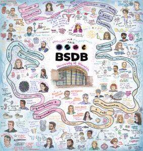
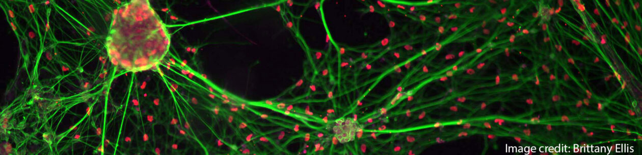
Illustration by Alex Neaverson, University of Cambridge
As members of the British Society for Developmental Biology, its annual meetings are always a highlight. This year’s ‘from Molecules to Morphogenesis’ meeting was held at University of Warwick in March, bringing together an inspiring range of talks spanning classical and modern developmental biology as well as a wide range of model systems.
Day 1: Seven Decades of Developmental Biology
On the first day we were all welcomed by a heartwarming introduction from Marysia Placzek (BSDB chair) talking about the history of the BSDB and the pioneer work of researchers that have shaped this society since the beginning, such as Philip Ingham who was present in the audience. The first session thus commemorated Seven Decades of Developmental Biology with a great line up of invited speakers highlighting the power of revisiting classical developmental questions with new technologies.
Starting with Denis Duboule remarkably showing in vitro embryo models retaining axis elongation and A-P polarity despite removing Hox gene function but showing defects in endodermal and mesodermal fate. Ruth Lehmann talked about the role of Nanos in early specification and maintenance of the Primordial Germ Cell (PGC) program in Drosophila embryos. Cliff Tabin added an evolutionary perspective on duck syrinx formation and morphological asymmetry through different types of Shh gradients. Yun Xia gave a fascinating talk on using ‘assembloids’ to study the interplay of cell repertoires in kidney development and disease progression. After a short coffee break, Alicia Hidalgo spoke about Toll signalling in brain plasticity and degeneration in flies. A standout talk for me was Norbert Perrimon’s on a decade long effort (also in flies!) to lay a framework of metabolic crosstalk between growing tumours and the host tissues that result in cachexia, a complex wasting syndrome. Andy McMahon described the underlying principles to direct iPSCs and generate human kidney organoids.
The last talk of the day was Valentina Lorenzi from the Wellcome Sanger Institute, who received the Beddington Medal for her impressive PhD work characterising spatiotemporal developmental trajectories of the human reproductive tract using cutting-edge technologies, but also for her work advancing and communicating women’s health as the former president of the Cambridge Femtech Society and through a collaborative zine called ‘Pelvic Matters’.
The Annual General Meeting followed, where all BSDB members get to hear from the committee what the society has been doing for the past year. All members also vote on changes to the constitution and on new committee members. The day ended with welcome drinks and dinner to encourage networking across all attendees.
Day 2: Cell Identity & Gene Regulation, Patterning & Morphogenesis
The first session of day two on Cell Identity and Gene Regulation, chaired by Vicki Metzis, began with Edith Heard on the dynamics of X-chromosome inactivation (XCI), followed by two short talks from Amruta Vasudevan, examining a Wnt/Nodal/Notch temporal module underlying symmetry breaking in mouse gastruloids, and Oliver Davis, highlighting the role of FoxG1 in regulating chromatin accessibility and neural fate in cerebral organoids. Joshua Brickman talked about using in vitro models to study chromatin remodelling and Sox2 activity. Following a short break and snacks, the session continued with Hilary Ashe, discussing ribosomal pausing and protein synthesis control in Drosophila embryos, and two more short talks. Christos Kalaitzis on the Hox code mediating regional diversity of vagal neural crest cells, and Daniel Goszczynski on dual developmental origins of mammalian PGCs.
There were also two rounds of flash talks from poster presenters, spanning varied topics including nuclear mechanosensing, transgenic avian lines, cell size scaling, cell morphology and more. During lunch time, the poster session provided a fantastic opportunity for delegates to dive deeper into the science, spark new conversations, and discover exciting work being done across the community. With packed rooms, lively discussions, and an exceptionally high standard of posters, the poster sessions were a highlight of the meeting.
The afternoon session covered Patterning & Morphogenesis under the lens of model organism diversity, chaired by Shankar Srinivas. Ranging from Drosophila to Arabidopsis, this was a unique opportunity to learn about how very distinct systems develop and shape their body plan or organs (e.g. mouse heart, fly blastoderm and epithelium, chick and shark telencephalon, mayfly eye, marsupial trunk, plant roots!). Several talks also highlighted the question of developmental timing. Kenzo Ivanovitch showed how early versus late primitive streak progenitors make distinct parts of the heart. Sergio Menchero from the Crick Institute gave the Dennis Summerbell Award lecture, showcasing his study on temporal diversity in marsupials’ developmental programs and how they prioritise differentiation of necessary structures for their survival. Dana Fakhreddine presented her work identifying heterochronic differences in molecular events shaping the distinct telencephalon identities. Erik Clark refined the classical morphogen-gradient models by adding a temporal lock mechanism to explain how striped expression is appearing in the fly embryo. Lastly, Bert De Rybel presented work from his lab on Arabidopsis root meristem, introducing the idea of developmental timers during development and aging.
After dinner two medal winners were announced. First, the Wolpert Medal was awarded to Prof. Neil Vargesson from U. of Aberdeen to recognise his outstanding contributions to our understanding of chemically induced birth differences for the past 20 years. Neil’s work has led to changes in health policies and help for affected families in the UK and abroad. His commitment to public engagement through numerous outreach activities has raised awareness of developmental biology and medicine safety. Next, the Waddington Medal lecture by this year’s awardee, James Briscoe from the Crick Institute. James’ fundamental discoveries have shaped our understanding of how morphogens work. He has also promoted developmental biology through numerous roles, including as Director and then Editor-in-Chief of the journal ‘Development’ from the Company of Biologists.
To end the evening, we had a Student/Postdoc ECR social event that lived up to expectations in every way. A science-themed pub quiz drew 120 attendees into teams that brought their competitive best. The questions ranged from easy to tricky and delightfully niche, a favourite was ‘when dealing with chick development, what does YSL stand for?’ and no, the answer is not Yves Saint Laurent but Yolk Syncytial Layer. Spirits were high and competition was tough, but one team came on top: ‘Shut up nurds!’ (a nod to Joshua Brickman’s talk). Perhaps the most creative team name came second, ‘Bad Scientists Doing Biology’ or, of course, B.S.D.B., and in third place we had the ‘Mighty-Chondria’ team. A great way to end day two!
Day 3: Cell Communication & Fate, Organogenesis & Regeneration, Conference Dinner
Wednesday morning started with the Cell Communication & Fate session, chaired by David Turner. This series of talks opened with Sally Lowell summarising elegant work from her lab generating new tools to track the neighbourhood of a cell and discussed how these tools can be applied to diverse model systems. Following this, a series of fascinating talks covered distinct systems where cell fate is controlled by intrinsic mechanisms, such as post-transcriptional modifications shaping neural crest fate as presented by Lara Busby, and extracellular cues provided by the environment, such as mechanical cues generated by neighbouring cells. On this topic, Stefan Harmansa presented his work on how mechanical forces shape epithelium architecture in the drosophila wing disk by combining experimental and modelling approaches. Vikas Trivedi shared recent work from his lab investigating the role of temperature on the shape of the zebrafish epithelial layer during gastrulation. Nine flash-talks concluded this session by covering a diversity of questions, ranging from the impact of metabolic alterations to the role of cellular geometry.
After lunch and the second poster session of this meeting, we returned for the afternoon session on Organogenesis & Regeneration, chaired by Timothy Saunders. This session underscored how the study of diverse systems furthers our general understanding of morphological and regenerative processes. Beautiful work from Emily Noël focused on the importance of ECM asymmetry in shaping the developing zebrafish heart. Cristina Newnes on muscle development in Drosophila embryos, showed the power of established model systems. Meanwhile, the comparative analysis of muscle development in zebrafish versus sharks from Peter Currie’s lab, the study of head scale formation in diverse reptiles by Rory Cooper, and the in vitro generation of hindlimb progenitors from hPSCs by Sude Uyulgan, all made clear how much there is to learn from studying novel or less conventional systems.
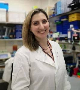
The Marie Johansson Prize was introduced for the first time by Corinne Houart, in memory of an outstanding Postdoc in her lab to recognise leading ECR researchers contributing to developmental biology. The first recipient of this prize is Giulia Boezio from the Crick Institute for her exceptional work on spinal cord patterning and establishing new techniques in complex embryonic tissues in vivo, as well as her contributions to public engagement, mentoring, and network. The Tickle Medal was then awarded to Cynthia Andoniadou from KCL for her incredible work as a developmental endocrinologist on the pituitary gland, building the human pituitary atlas, and championing female scientists in the UK and internationally.
Ending the last full day, we had the long-awaited Conference Dinner, during which Marysia Placzek acknowledged and thanked the meeting organisers with a special gift for making this meeting happen. She then announced the poster prize runner ups and winners: Gareth Moore (£150) and Luke Simpson (£300) in the postdoc category, Achira Karunaratna (£150) and Noura Maziak in the student category. Noura won the big prize of an all-expenses-paid trip to attend the Society for Developmental Biology meeting in the US! The new members of the BSDB committee were also announced, congratulations and welcome to Paula Alexandre, Gi Fay (Geoffrey) Mok, and Teresa Rayon. Finally, it wouldn’t be a BSDB meeting without lots of scientists cheering and dancing the night away!
Day 4: Human Development & Disease
Our final session of the conference mercifully started at 9.30, giving us extra time to recover after the night before. This session was all about Human Development & Disease, chaired by Anahí Binagui-Casas. Sarah Teichmann showed the incredible efforts of the Human Developmental Cell Atlas, which has mapped >450 cell types across pre- and postnatal development, an invaluable resource for the community. This work also highlighted an underlying theme across the conference that many progenitor populations and lineage commitments emerge earlier in development than previously anticipated. Presentations from Lila Allou, Teresa P. Silva, and Lorenz Studer also made clear, in a sometimes-poignant manner, how garnering a deeper understanding of developmental processes such as organogenesis and axis formation can help us to better understand how such processes change in the context of congenital diseases. Kathy Niakan shared the molecular mechanisms regulating the first cell fate decisions in human embryos but also made a noteworthy remark about supporting model organisms, highlighting that “we wouldn’t be able to know what to look for (in humans) if it wasn’t for work in other organisms”.
Overall, this was an enjoyable and memorable meeting thanks to all the attendees, speakers, organisers, and everyone in the community coming together to share their passion for developmental biology. Looking forward to next year’s BSDB meeting in Edinburgh!
P.S. Check the BSDB social media to relive the highlights of the 2026 meeting! @bsdb.socials on Instagram and TikTok
Irene Amblard
Thamarailingam Athilingam
Alejandra Guzman-Herrera
Dimitra Mouzourou
Andrew Plygawko
 The
The 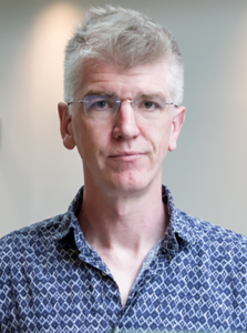
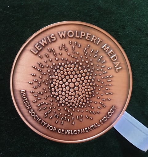
 We are very happy to announce that this year’s winner of the BSDB Wolpert medal is Prof. Neil Vargesson from the University of Aberdeen.