Welcome from our new chair

Dear members of the BSDB – I have recently taken over as President/Chairman of our society from the wonderful Ottoline Leyser and will try my hardest to do as good a job as she in advocating and looking after the interests of developmental biologists in the UK. Please let me know urgent things that you think I need to know about and that you’d like me to act upon.
I have heard that the EDBC meeting in Alicante was a really great meeting, and am happy that many BSDB members were able to attend. I would have loved to have been at this first European Developmental Biology meeting for several years but couldn’t attend because I had tonnes of teaching that week. Please be sure to note what are the highlights of this meeting (as well as things that don’t work so well) because the BSDB is hosting the next one of these meetings in the UK in 2023 and we want to make it a big success. The meeting itself will be co-organised with other European developmental biology societies, and we look forward to working with them on this.
Hoping to meet with lots of you in Warwick next March at our annual Spring meeting. In the meantime good luck with all your science and here’s hoping that all your 3rd referees are kind ones.
Paul Martin
Upcoming meetings in 2020!
Registration for the Spring 2020 BSDB/Genetics society meeting is now live!
All details about this fantastic meeting can be found here. Please bookmark this page to keep up to date with plans for our annual meeting
Our Autumn 2020 meeting will be held jointly with the International Society for Differentiation. It will take place on 13-16 Sept 2020 in the beautiful location of Valletta, Malta and will be organised by Liz Robertson, Josh Brickman & Sally Lowell. BSDB Conference grants will be available for our members. We have a great line up of speakers for this meeting- details to be released soon.
Hello, Goodbye, Hello, Goodbye…
All members of the committee want to take this opportunity to thank Ottoline Leyser for her dedication and efforts during her time as the BSDB chair. Its been great having your help during our various transitions! We also welcome Paul into his new role!
In addition, we have had a change in the treasurer role this year, saying goodbye to Chris Thompson and hello to Cynthia Andoniadou. A huge thanks to Chris for keeping our finances in check over the last five years! We also have Andy Oates leaving the committee this autumn, so thanks &y for your input and enthusiasm over the last five years.
Please keep an eye out for future nominations to join the committee and contribute the running of our society and the organisation of our meetings. We always strive to obtain a broader representation of our membership as possible, both in terms of geographical and model organism representation. So… if you are a plant developmental biologist- please stand up!
Waddington medal lecture 2019:
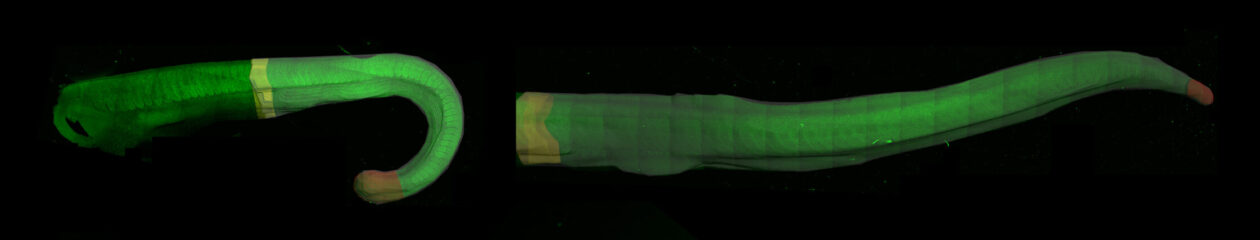
For those who couldn’t make it to the Spring lecture this year, or for those wanting to watch Kate Storey’s inspiring lecture again by following this link to our YouTube channel. There you will also find previous talks that we have recorded from our meetings

PlanS delayed by a year
In our Spring newsletter, we featured and article looking into how PlanS might impact society journals as it becomes implemented. It is an initiative set up by a consortium of research funders called cOAlition S and aims to rapidly promote the transition of publishes to a fully open access model. While this is widely supported by the community, there are concerns about how this will effect journals operating on a non-for-profit model, who re-insert funds gained from their journals back into the community. A key example for us as developmental biologists, is the Company of Biologists who generously provide funds to the BSDB and other academic societies, as well as supporting the community through the provision of travel grants, conference organisation and the maintenance of the Node (as well as many other things!). The decision of cOAlition S to delay implementation by a year will provide a welcome breathing space to publishes who are supportive of the underlying principles of open access, but nevertheless require more time to make the necessary changes to their publishing models.
“With effect from 2021, all scholarly publications on the results from research funded by public or private grants provided by national, regional and international research councils and funding bodies, must be published in Open Access Journals, on Open Access Platforms, or made immediately available through Open Access Repositories without embargo.”
cOAlition S
In addition to delaying implementation until 2021, the consortium have said that they won’t immediately place a cap on open access charges that journals will be able to charge to their authors. In addition, they have changed the rules around hybrid journals, in the hope that this will allow partly paywalled journals to have an easier transition to becoming fully open access.