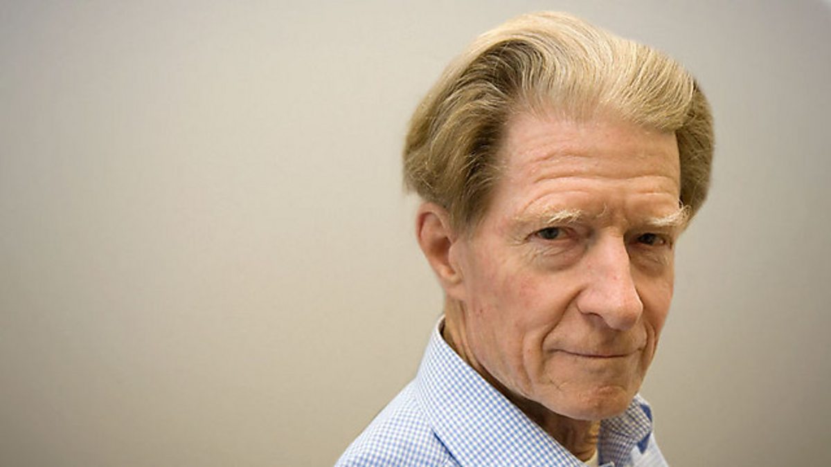
John Gurdon was an extraordinary man who had an immense impact on the whole field of Developmental Biology. He pioneered the concept that cell differentiation occurs by selective gene expression and not by irreversible loss of genetic information, a milestone in biology which he achieved by transplanting nuclei from embryonic cells, or specialised cells, into eggs whose nucleus he had destroyed. By-products of this work were animal cloning and Dolly the sheep. He also showed that the proportion of pluripotent cells decreases during development, but that some persist, providing early evidence of adult stem cells. He shared the Nobel Prize for Physiology or Medicine with Shinya Yamanaka in 2012.
John was an influential mentor to me. I first met him in 1964, when a demonstrator in a first-year practical class insisted that I should take my two-headed tadpole to his lab to show him. I was innocently unaware that John had retreated to his lab to evade dissatisfied students as he was sure the grafting experiment wouldn’t work. Fortunately, John remembered that first encounter two years later when I applied to become his graduate student. He remained a close colleague for over a third of a century and a good friend until he died, five days after his 92nd birthday.
John’s introduction to biology at Eton was notoriously unsuccessful. Not only was he ranked last in biology out of 250 pupils, but his biology teacher wrote: “He will not listen, but will insist on doing his work in his own way.” To his credit, this teacher inadvertently recognized one of John’s greatest strengths, an unswerving determination to pursue his own goals single mindedly, against all odds. The same school report added: “I believe he has ideas of becoming a scientist; on his present showing this is quite ridiculous.” Fortunately, after he gained First Class Honours in Zoology at Oxford, a Lecturer in Developmental Biology, Michail Fischberg disagreed and invited him to try transplanting nuclei in the frog Xenopus, building on a method for Rana recently published by Briggs and King and adapted by Fischberg and Tom Elsdale.
John’s efforts as a graduate student in Oxford resulted in two Nature papers that supported the concept that cell differentiation can occur without irreversible loss of genetic information. This concept is so firmly established now that it is difficult to remember that it continued to attract opposition until the late 1970s. In the course of this work, John pioneered the method of serial nuclear transplantation, which produced clones of genetically identical animals. The use of this concept, first in science fiction and now in agriculture has made it feel familiar, though at the time it was entirely novel. John invented animal cloning.
Using serial nuclear transfers, John showed that nuclei from visibly differentiated intestinal epithelium cells from swimming tadpoles could support the development of fertile adult frogs. This same approach also demonstrated that the proportion of nuclei that were pluripotent after transplantation decreased during embryonic development. As a graduate student, I attempted to extend nuclear transfers to nuclei from adult donor cells, instead of embryonic donors. Together, John and I demonstrated that nuclei from a range of adult cells including lung, kidney, heart and skin could programme enucleated eggs to develop as far as the feeding tadpole stage. To test adult skin cells, we cultured skin explants and saw outgrowths of undifferentiated cells that then synthesized immunoreactive keratin. Nuclei from these keratinizing cells supported development as efficiently as other adult cells, emphasising that differentiation could occur without irreversible loss of genes. It also demonstrated that at least some of the cells in a differentiated adult tissue were highly pluripotent. Nowadays we would refer to them as adult stem cells.
John’s momentous scientific discoveries go much further. By transplanting nuclei into foreign cytoplasm he showed that cytoplasmic factors drive the cell cycle and gene expression. He demonstrated the existence of a cytoplasmic factor that induces mitosis, later called MPF, and another that induces DNA synthesis. Both were shown by others to consist of cyclins and cyclin dependent kinases, but it was John who discovered their existence. He also demonstrated that the cytoplasm determines patterns of gene activity of transplanted nuclei and simultaneously that targeting of proteins to the nucleus from the cytoplasm is specified by information in the mature structure of the proteins.
As John Gurdon’s career progressed he used molecular techniques to understand development and differentiation. He microinjected purified macromolecules into Xenopus eggs to demonstrate and analyse DNA replication, transcription of cloned DNA into RNA and mRNA translation into protein. This allowed others to exploit these systems to isolate the genes encoding, for example, interferon as well as neurotransmitters and their receptors.
More recently John turned his attention to analyse mechanisms of cell interaction and intercellular signaling during development. This led him to discover a community effect, which defines the demarcation between cell populations and establishes uniformity within them. He showed it was mediated by threshold responses to signal factor concentrations and that a single embryonic cell can follow several different cell fates depending on the concentration of a single identified signal factor. He also elucidated mechanisms by which cells perceive their position in a concentration gradient of signaling factors and respond accordingly. Later John’s work came full circle as he elucidated the chromatin changes that accompany and mediate the reprogramming of transplanted nuclei.
Taken together John Gurdon’s contributions amount to an extraordinary body of major discoveries in cell biology and development. His achievements were recognized in many ways. He gave three of The Royal Society’s named lectures and received their two most prestigious medals, the Royal Medal and the Copley Medal. He was Knighted in 1995 and he received honours from many countries including Belgium, France, Germany, Israel, Italy, Sweden, Switzerland, Japan and the USA. He received Honorary Degrees from both Oxford and Cambridge amongst many others. He also had the rare distinction of having a major institute named after him during his working life-time. The Wellcome Trust, Cancer Research UK, Gurdon Institute was founded by John together with Sir Martin Evans, Chris Wylie, Janet Heasman, Michael Akam and myself, joined soon after by Azim Surani and Dame Anne McLaren.
Although he disliked University administration, John served on many national and international committees and boards. For example, he was President of the International Society for Developmental Biology and he chaired the Company of Biologists for 10 years. Surprisingly, he became Master of Magdalene College Cambridge, where his wife Jean rose brilliantly to the challenges that college life imposed.
 |
| Sketch, courtesy of Virginia E. Papaioannou |
A second sphere in which John Gurdon excelled was his exceptional standard of undergraduate teaching. In the light of all this, you may have an image of a highly focused and perhaps rather one-dimensional individual. Nothing could be further from the truth. In addition to world leading cell and developmental biology, John Gurdon found time to represent Oxford University at squash and to be a Junior British Squash Doubles Champion, to represent Oxford University at skiing, to drive very fast cars, to become an expert on butterflies and moths, alpine botany and desert botany and to give the rest of his colleagues glimpses of infuriating abilities in all sorts of other areas including tennis, croquet, skating and mountaineering. Working closely with John for a third of a century had one serious disadvantage. John was eleven years older than me, but a woman giving directions for finding a room at an institute retreat pointed to John and told me to follow that young man over there.
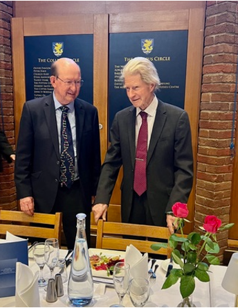 |
| Ron Laskey and John Gurdon, courtesy of John’s daughter, Aurea |
John had a mischievous sense of humour and it would be possible to list many more things about him, such as his single-handed support of McVitie’s biscuit sales, especially when travelling to countries whose food he disliked, – notably France! This led to his remarkable discovery that excess consumption of McVitie’s biscuits is the secret to hair retention; I should have started eating them decades ago. Instead I shall conclude by adding my amazement at John’s powers of endurance in tolerating one close colleague for a third of a century and even managing to maintain a harmonious relationship while he was my Chairman and simultaneously, I was his Director, a tangle which many would find difficult to unravel, but with John the positives always outweighed the negatives. I and many others will remain deeply grateful to John and I am sure that his impact will endure, not just in his discoveries, but also in the work of his many protégés, of whom I am grateful to be one.
Ron Laskey
Emeritus Charles Darwin Professor of Animal Embryology
University of Cambridge
 The
The 
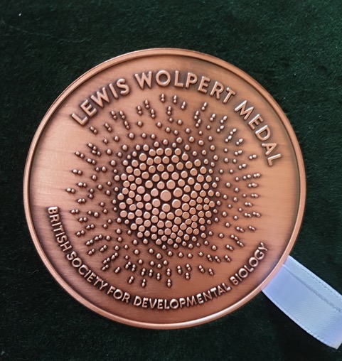
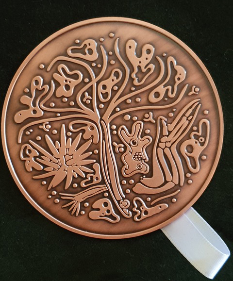 We are very happy to announce that this year’s winner of the BSDB Wolpert medal is Prof. Neil Vargesson from the University of Aberdeen.